Article
Welcome to EyeICare
Article #2 : Eye injury
2-March-2015
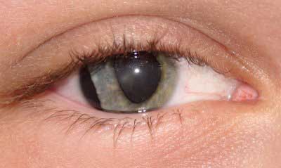
Physical or chemical injuries of the eye can be a serious threat to vision if not treated appropriately and in a timely fashion. The most obvious presentation of ocular (eye) injuries is redness and pain of the affected eyes. This is not, however, universally true, as tiny metallic projectiles may cause neither symptom. Tiny metallic projectiles should be suspected when a patient reports metal on metal contact, such as with hammering a metal surface. Intraocular foreign bodies do not cause pain because of the lack of nerve endings in the vitreous humour and retina that can transmit pain sensations. As such, general or emergency room doctors should refer cases involving the posterior segment of the eye or intraocular foreign bodies to an ophthalmologist. Ideally, ointment would not be used when referring to an ophthalmologist, since it diminishes the ability to carry out a thorough eye examination. Flicking sand, flying pieces of wood, metal, glass, stone and other material are notorious for causing much of the eye trauma. Sporting balls such as cricket ball, lawn tennis ball, squash ball, shuttle cock (from Badminton) and other high speed flying objects can strike the eye. The eye is also susceptible to blunt trauma in a fistfight. The games of young children such as bow-and-arrows, bb guns and firecrackers can lead to eye trauma. Road traffic accidents (RTAs) with head and facial trauma may also have an eye injury - these are usually severe in nature with multiple lacerations, shards of glasses embedded in tissues, orbital fractures, severe hematoma and penetrating open-globe injuries with prolapse of eye contents. Other causes of intraocular trauma may arise from workplace tools or even common household implements. Depending on the type of ocular injury, either a pressure patch or shield patch should be applied. Up until circa 1987, pressure patches were the preferred method of treatment for corneal abrasions in non-contact lens wearers; Multiple controlled studies conducted by accredited organizations such as the American Academy of Ophthalmology have shown that pressure patching is of little or no value in healing corneal abrasions and is actually detrimental to healing in some cases. Pressure patching should never be used on an individual presenting with a corneal abrasion who has a history of contact lens wear. In this circumstance, a virulent infection caused by the bacterium Pseudomonas aeruginosa is at a clearly delineated increased risk for occurrence. These infections can cause blindness within 24 – 48 hours and there is a possibility that the infection can move into the peri-orbital socket, resulting in the need for evisceration of the eyeball. In rare cases, the infection can enter the brain and cause death to the patient. In cases of globe penetration, pressure patches should never be applied, and instead a shield patch should be applied that protects the eye without applying any pressure. If a shield patch is applied to one eye, the other eye should also be patched due to eye movement. If the uninjured eye moves, the injured eye will also move involuntarily possibly causing more damage.
Article #1 : Arthropod eye
1-March-2015
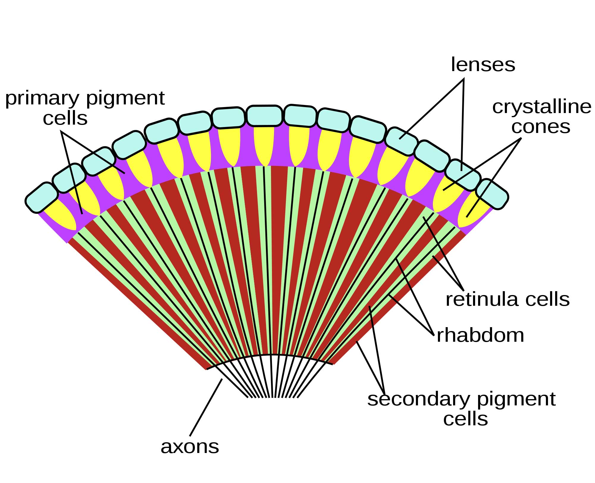
The eyes of trilobites came in three forms. The holochroal eye, the most common and most primitive, consisted of many small lenses, between 100-15000, covered by a single corneal membrane. This was the most ancient kind of eye. This eye morphology was found in the Cambrian trilobites (the earliest) and survived until the Permian extinction. The more complex schizochroal eye was found only in one sub-order of trilobite, the Phacopina (Ordovician-Silurian). It has no modern counterpart. The eye has up to 700 larger lenses with individual sclera separating each lens. Each lens has a cornea. Schizochroal eyes developed from holochroal ones and were more powerful with overlapping visual fields. They were particularly useful for nocturnal vision and possibly for colour and depth analysis. The lenses of the eye were constructed from single calcite crystals. Early schizochroal eye designs were rather haphazard and irregular, though constrained by the geometrical requirements of packing identical sized lenses on a curved surface. Later designs used graduating lens sizes. The third eye morphology of trilobites, called the abathochroal, was found only within the Eodiscina. This morphology consisted of up to 70 much smaller lenses. The cornea separated each lens, and the sclera on each lens terminated on top of each cornea. The eye morphology of trilobites is useful for determining their mode of life, and can function as palaeoenvironmental indicators. The horseshoe crab has traditionally been used in investigations into the eye, because it has relatively large ommatidia with large nerve fibres (making them easy to experiment on). It also falls in the stem group of the chelicerates; its eyes are believed to represent the ancestral condition because they have changed so little over evolutionary time. Indeed the horseshoe crabs are often considered to be living fossils. Most other living chelicerates have lost their lateral compound eyes, evolving simple eyes in their place. Horseshoe crabs have two large compound eyes on the sides of its head. An additional simple eye is positioned at the rear of each of these structures. In addition to these obvious structures, it also has two smaller ocelli situated in the middle-front of its carapace, which may superficially be mistaken for nostrils. A further simple eye is located beneath these, on the underside of the carapace. A further pair of simple eyes are positioned just in front of the mouth.[16] The simple eyes are probably important during the embryonic or larval stages of the organism, with the compound eyes and median ocelli becoming the dominant sight organisms during adulthood. These ocelli are less complex, and probably less derived, than those of the mandibulata. Unlike the trilobites', the compound eyes of horseshoe crabs are triangular in shape; they also have a generative region at their base, but this elongates with time. Hence the one ommatidium at the apex of the triangle was the original "eye" of the larval organism, with subsequent rows added as the organism grew.